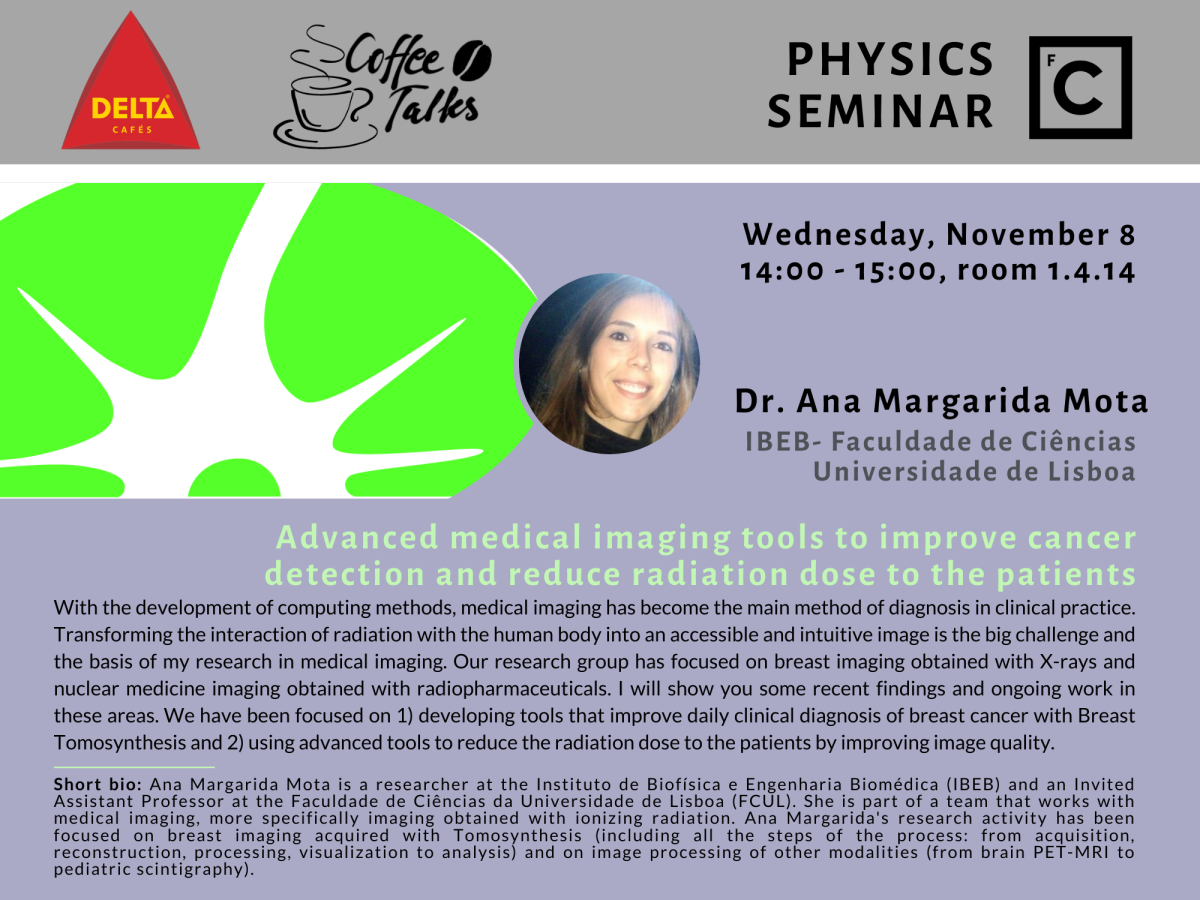
Por Ana Margarida Mota (IBEB, FCUL).
With the development of computing methods, medical imaging has become the main method of diagnosis in clinical practice. Transforming the interaction of radiation with the human body into an accessible and intuitive image is the big challenge and the basis of my research in medical imaging. Our research group has focused on breast imaging obtained with X-rays and nuclear medicine imaging obtained with radiopharmaceuticals. I will show you some recent findings and ongoing work in these areas. We have been focused on 1) developing tools that improve daily clinical diagnosis of breast cancer with Breast Tomosynthesis and 2) using advanced tools to reduce the radiation dose to the patients by improving image quality.
Short bio: Ana Margarida Mota is a researcher at the Instituto de Biofísica e Engenharia Biomédica (IBEB) and an Invited Assistant Professor at the Faculdade de Ciências da Universidade de Lisboa (FCUL). She is part of a team that works with medical imaging, more specifically imaging obtained with ionizing radiation. Ana Margarida's research activity has been focused on breast imaging acquired with Tomosynthesis (including all the steps of the process: from acquisition, reconstruction, processing, visualization to analysis) and on image processing of other modalities (from brain PET-MRI to pediatric scintigraphy).


